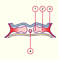
7.2 The trilaminar germ disk (3rd week)


The intraembryonic coelom


The intraembryonic coelom first appears in the lateral plate mesoderm in the form of several isolated vacuoles. During the lateral unfolding of the embryo in the 4th week these vacuoles fuse and form a U-shaped cavity: the intraembryonic coelom. In the beginning a connection exists between the intra- and extraembryonic coeloms.
With the progress of the unfolding, though, the merging of the ectoblast layers along the medial line has the effect that the intraembryonic coelom is separated from the extraembryonic one and remains enclosed in the lateral mesoblast.
Fig. 19 - Mesoderm before the
vacuoles form on roughly the 20th day
 Legend

1
2
3
4
Paraxial mesoderm
Intermediate mesoderm
Lateral plate mesoderm
Chordal process

Fig. 19
Transverse section through a 20-day-old embryo. The separation into the paraxial, intermediate mesoderm and the lateral plate is shown.


Fig. 20 - Coelomic vacuoles at roughly
the 23rd day
Fig. 21 - Coelomic cavity at roughly
the 25th day
 Legend

1
2
3
4
5
6
7
Lateral plate mesoderm
Intermediate mesoderm
Paraxial mesoderm
Neural groove
Coelomic vacuoles
Intraembryonic coelom
Extraembryonic coelom
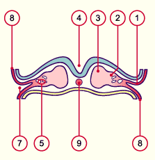

8
9
10
11
12
Extraembryonic mesoblast
Notochord
Splanchnopleure with endoderm
Somatopleure with ectoderm
Dorsal aorta (paired)

Fig. 20
After the 23rd day coelomic vacuoles form in the lateral plate.

Fig. 21
On the 25th day the intraembryonic coelom of the lateral plate divides the mesoderm into somatopleural und splanchnopleural mesoderm.


Previous page | Next page