|

|
|
|
7.2 The trilaminar germ disk (3rd week)
|
|
|
Induction of the neural plate - neurulation
|
|
|
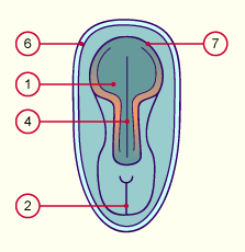
| Fig. 22 - Neural plate: 19 – 23rd day |
|
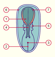
Fig. 23 - Neural plate at roughly
the 25th day |
|
Legend |

1
2
3
4
|
Neural plate
Primitive streak
Primitive nodes
Neural groove |
|
|
|

5
6
7
|
Somites
Cut section of the amnion
Neural folds |
|
|
|
Fig. 22
With the appearance of the neural plate on the 19th day, the development of the future nervous system begins.
Fig. 23
The cranial end the neural plate is wider and encloses the region where the brain will arise.
The caudal end is narrower; there the spinal cord will form.
|
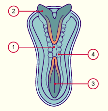
Fig. 24 - The neural tube at roughly
the 28th day |
|
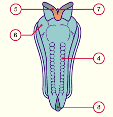
Fig. 25 - The neural tube at roughly
the 29th day |
|
Legend |

1
2
3
4
|
Neural tube
Neural fold
Neural groove
Somites |
|
|
|

5
6
7
8
|
Neural crest
Protrusion of the pericardium
Cranial neuropore
Caudal neuropore |
|
|
|
Fig. 24
In the course of the 3rd week the edges of the neural plate rise and form two folds that bound the neural groove.
Fig. 25
The closure of the neural tube begins in the cervical area (in the middle of the embryo) and spreads from there in the cranial and caudal directions.
|
|
The neural crest cells  9 9 form, so to speak, a 4th embryonic germinal layer. This contains a partial segmentation that contributes to the formation of the peripheral nervous system (neurons and glia cells of the sympathetic, parasympathetic and sensory nervous systems). form, so to speak, a 4th embryonic germinal layer. This contains a partial segmentation that contributes to the formation of the peripheral nervous system (neurons and glia cells of the sympathetic, parasympathetic and sensory nervous systems).
The neural crest cells are distinguished by a great migrating ability and phenotypic heterogeneity, since numerous and various differentiated cell types will arise from them. Not only do the nerve and glia cells mentioned above arise from the neural crest, the epidermal pigment cells (melanocytes), the calcitonin cells of the thyroid gland, the cells of the adrenal medulla and some components of the skeletal and connective tissue in the head area are also generated from them.
|
|
|
| Fig. 26 - The forming neural crest (neural plate stage) |
|
Legend |

A
B
1
2
3
|
Neural plate stage
Neural groove stage
Epiblast
Neural groove
Neural crest |
|
|
|
Fig. 26
The beginning of neurulation in the cervical region.
The neural groove forms. The cells of the future neural crest are in orange. The arrows show the direction of the lateral folding.
|
|
|

