|

|
|
 |
19.1 Face and upper foregut
|
|
|
|
The tongue begins to form at approximately the same time as the palates. It extends from various protuberances on the pharynx floor. Already at the time of the medial fusion of the first (mandibular) and second (hyoid) pharyngeal arches a medial protuberance, the tuberculum impar, appears on the lower edge of the mandibular arch. To the left and right of it two further swellings form, the lateral lingual prominences.
|
|
|
|
|
|
These three swellings extend from the mandibular arch and later form the anterior 2/3 of the tongue. Thus this part the tongue consists of both ectodermic and endodermic portions. At the caudal end of the tuberculum impar arises the primordium (or anlage) of the thyroid (stage 10, ca. 28 days  10 10 ) as an unpaired ventral endodermic bud. In its further development it migrates in front of the larynx and, at the invagination site at the tongue base, leaves behind the foramen cecum. The posterior third is formed from the material of the fusion site of the 2nd and 3rd pharyngeal arches, the copula, and a small portion of the 4th pharyngeal arch. It only consists of endodermic parts. The third pharyngeal arch probably grows over the second one so that in the end the second one has no mesenchymal contribution to tongue formation. Between the anterior two-thirds and the posterior third the terminal sulcus is found. The tongue is delimited caudally by the hypopharyngeal eminence that marks the entrance into the trachea (laryngeal inlet). ) as an unpaired ventral endodermic bud. In its further development it migrates in front of the larynx and, at the invagination site at the tongue base, leaves behind the foramen cecum. The posterior third is formed from the material of the fusion site of the 2nd and 3rd pharyngeal arches, the copula, and a small portion of the 4th pharyngeal arch. It only consists of endodermic parts. The third pharyngeal arch probably grows over the second one so that in the end the second one has no mesenchymal contribution to tongue formation. Between the anterior two-thirds and the posterior third the terminal sulcus is found. The tongue is delimited caudally by the hypopharyngeal eminence that marks the entrance into the trachea (laryngeal inlet). |
|
|
|
Lingual musculature stems from muscle cells that immigrate from the occipital somites into the tongue. The emigrating predecessor cells follow the hypoglossal nerve (CN 12). |
|
Fig. 10 - Embryo in stage 14
ca. 33 days |
|
Legend |
|
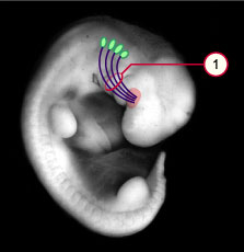
1
|
Hypoglossal tongue cord |
|
|
|
Fig. 10
The occipital somites are shown in green. The tongue receives material from all 4 occipital somites for building up the musculature. The connective tissue and the myoblasts together form a hypoglossal cord.
|
|
Development of the tongue involves several pharyngeal arches. This is also evidenced in their innervation that at the first moment seems relatively complex.
|
|
Fig. 11 - Innervation of the tongue |
|
Legend |
|
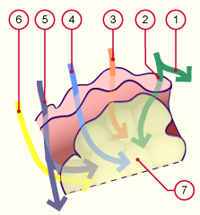
| 1 |
Maxillary nerve (CN V2) |
2
|
Lingual nerve
(mandibular nerve, CN V3) |
3
4
5
6
7 |
Chorda tympani
(facial nerve, CN VII)
Glossopharyngeal nerve (CN IX)
Vagus nerve (CN X)
Hypoglossal nerve (CN XII)
Tongue |
|
|
|
Fig. 11
Cerebral nerves of the pharyngeal arches grow into the tongue and provide it with motor, sensitive, and sensorial innervation.
|
| Sensitivity: (touch) |
| Anterior two-thirds of the tongue |
1. Pharyngeal arch |
N. lingualis (CN V3) |
| Posterior third of the tongue |
3. Pharyngeal arch |
N. glossopharyngeus (CN IX) |
| Tongue base |
4. Pharyngeal arch |
N. vagus (CN X) |
| Sensorial: (taste) |
| Anterior two-thirds of the tongue |
2. Pharyngeal arch |
Chorda tympani (CN VII) |
| Posterior third of the tongue |
3. Pharyngeal arch |
N. glossopharyngeus (CN IX) |
| 4. Pharyngeal arch |
N. vagus (CN X) |
| Motor: (movement) |
| Entire musculature |
6. Pharyngeal arch |
N. hypoglossus (CN XII) |
|
|