|

|
|
|
19.1 Face and upper foregut
|
|
|
Definitive pharynx: Derivatives of the first and second pharyngeal arches
|
|
|
|
The embryonic pharynx represents the cranial section of the foregut. On the side and ventrally it is surrounded by the pharyngeal arches. The definitive pharynx consists of three parts:
- Nasopharynx
- Oropharynx
- Laryngopharynx
The differentiated oral cavity now no longer counts as part of the definitive pharynx. An important property of a developing definitive pharynx is the series of pharyngeal arches and pouches that are associated with the formation of the thyroid, parathyroid, the tonsils, middle ear, the thymus, the epithelial bodies and the trachea. It is to be noted that, except the tonsils and the anlage of the middle ear, which always retain a connection to the pharynx, these structures have a close relationship with the developing heart. This explains the fact that, with the progressing descent of the heart, all of these structures come to be located in the lower neck or thorax region.
|
|
|
|
The primordium of the thyroid (stage 10, ca. 28 days,  10 10 ) forms as a median thickening on the floor between the first and second pharyngeal arches. Soon a bilobular sacculation of the endoderm develops out of it that remains connected with the mouth floor by the thyreoglossal duct. This also gets thinner and thinner and, with the further descent, the epithelial cord disintegrates and the thyroid loses its contact with the mouth floor. What remains is the foramen cecum. ) forms as a median thickening on the floor between the first and second pharyngeal arches. Soon a bilobular sacculation of the endoderm develops out of it that remains connected with the mouth floor by the thyreoglossal duct. This also gets thinner and thinner and, with the further descent, the epithelial cord disintegrates and the thyroid loses its contact with the mouth floor. What remains is the foramen cecum.
(see the overview about the descent of the derivatives of the third and fourth pharyngeal pouches)
|
|
|
|
On the side, between the first and second pharyngeal arches the first pharyngeal pouch forms on the inside in parallel with the first pharyngeal cleft that is visible on the outside. This pharyngeal pouch lengthens but always remains in connection with the throat (tubotympanic recess for the middle ear cavity and the Eustachian tube). On the one side, its dead-end gets a close relationship thereby with the inner ear and, on the other, it forms the anlage material for the eardrum (tympanon) with the bordering ectoderm of the first pharyngeal cleft.
(see the overview about the descent of the derivatives of the third and fourth pharyngeal pouches)
|
|
|
|
At the beginning of the digestive tract, at the transition between the oral and nasal cavities in the oropharynx, there is a large collection of lymphatic tissue. This tonsillar ring (Waldeyer's tonsillar ring) is composed out of several lymphoreticular organs. A few lymph follicles can be found scattered in the whole region of the pharyngeal mucosa, especially around the posterior nasal orifice, on the isthmus of the fauces, the piriform recess and laryngeal inlet. Accumulations of lymphatic tissue, though, are found especially in the region of the tonsils. Four lymphoepithelial organs can be distinguished:
- pharyngeal tonsil
- tubal tonsil
- palatine tonsil
- lingual tonsil
These lymphatic organs of the tonsillar ring make possible an initial defense against infection at the beginning of the digestion process and respiration. These lymphatic organs arise only in the early fetal period since up till birth no defense against infection needs to take place.
The pharyngeal tonsil arises in the roof of the nasopharynx. The mucosa epithelium (here respiratory epithelium) is opened up in folds thereby and, under it, lymphatic tissue accumulates. The portion of the lymphatic tissue around the oral tubal pharynx is called the tubal tonsil.
In the young fetus, the palatine tonsil forms on both sides in the tonsillar sinus as the remainder of the 2nd pharyngeal pouch. The epithelium thickens and under it a mesenchymal accumulation forms from material of the second pharyngeal arch. At the same time, the blood and lymph vessel system also gets denser in this region. Endodermal epithelium cords begin to grow into the underlying mesodermal blastema and later form the cryptae of the first and second order.
|
|
|
The lingual tonsil forms on the tongue floor in the region of the terminal sulcus. As in the palatine tonsil the mucosa invades into the depths with short canals and forms tonsillar crypts. These become bounded by lymphatic tissue that is surrounded by a connective tissue capsule. On the surface their openings are crater-shaped depressions in protuberances of the mucosa, the so called lenticular papillae of the tongue base.
(see the overview about the descent of the derivatives of the third and fourth pharyngeal pouches) |
|
Fig. 16 - Base of the tongue with
terminal sulcus |
|
Legend |
|
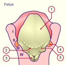
1
2
3
4
5
I
II
III
IV |
Tongue
Terminal sulcus
Arythenoid prominence
Palatine tonsil
Lenticular papillae
Pharyngeal arch
Pharyngeal arch
Pharyngeal arch
Pharyngeal arch |
|
|
|
Fig. 16
The palatine tonsils and the lenticular papillae are part of Waldeyer's pharyngeal ring.
|
|
|