|

|
|
|
19.1 Face and upper foregut
|
|
|
|
One subdivides the human denture into four jaw quadrants. In each one 5 deciduous (or milk) teeth and later 8 permanent teeth form. Quadrants 1-4 are numbered clockwise (first number); the teeth from mesial towards distal in each of the four quadrants (second number).
|
|
|
|
right
|
mesial
|
left
|
|
18 17 16
|
15 14 13 12 11 21 22 23 24 25 |
26 27 28
|
|
48 47 46
|
45 44 43 42 41 31 32 33 34 35 |
36 37 38
|
|
The development of the teeth begins in the late embryonic period (stage 18, ca. 44 days,  18 18 ) with the formation of an epithelial lamella parallel to the lip edge. This ridge, the labiodental lamina, later forms a furrow (the labiodental sulcus), out of which the oral vestibule arises. Dental development also begins through a ridge-shaped thickening on this labiodental lamina that is oriented towards the oral cavity. A U-shaped band, the dental lamina arises. ) with the formation of an epithelial lamella parallel to the lip edge. This ridge, the labiodental lamina, later forms a furrow (the labiodental sulcus), out of which the oral vestibule arises. Dental development also begins through a ridge-shaped thickening on this labiodental lamina that is oriented towards the oral cavity. A U-shaped band, the dental lamina arises.
Through interactions between neural crest cells and ectoderm 10 roundish teeth buds soon grow in the lower and upper jaws on the labial side of this dental lamina (early fetal period), which represent the primordia of the deciduous teeth. Somewhat later (ca. 16 weeks) small buds also form on the oral side. These are the early anlagen of the permanent teeth.
|
|
|
|
|
 |
| Each teeth bud has thus an ectodermal origin and is also called an enamel organ. On the inside, they surround a solidified mesenchyma of neurectodermal origin that forms the dental pulp. The edges of the enamel organ grow faster than the middle part, thus forming a dental bell that arises from the tooth bud via a cap-shaped stage. The mesenchyma around the whole tooth bud also condenses and forms the dental follicle. Out of it arises the periodontium and the cement of the tooth root. |
|
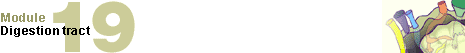
Fig. 12 - Development of a tooth
from dental bud to dental bell stage |
|
Legend |
|
1
2
|
Cap-shaped tooth bud
Tooth bell
|
|
|
|
Fig. 12
The tooth bud arises as a bud from the U-shaped dental lamina. The edges grow faster than the interior so that soon a bell is formed.
|
|
In an enamel organ one distinguishes between an outer and an inner enamel epithelium. Between them is found the enamel pulp. The enamel epithelium is nourished from the outside by a capillary network while the enamel pulp always remains vessel free. The ameloblast layer, which forms out of the inner enamel epithelium, produces the enamel - in the form of enamel prisms - in the direction of the tooth pulp.
Immediately next to the inner enamel epithelium the mesenchyma that lies below it organizes itself into an epithelial layer, the odontoblast layer, which excretes predentin towards the outside. Dentin then arises through deposits of calcium salts in the predentin.
|
|
|
Synonyms
|
|
Ameloblast
=
Adamantoblast
|
|
|
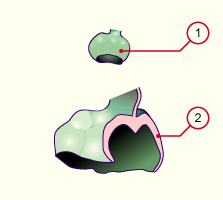
Fig. 13 - Anlage of a deciduous tooth
with its permanent tooth |
|
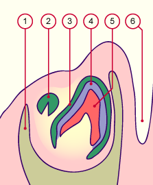
Fig. 14 - Structure of the enamel organ
before and after the beginning
of the enamel deposition |
|
Legend |
1
2
3
4
5
6
|
Mandible
Anlage of the permanent tooth
Enamel organ
Enamel
Dentin
Labiogingival sulcus |
|
|
|
1
2
3
4
5
6 |
Outer layer of the enamel organ
Enamel pulp
Inner layer of the enamel organ
(ameloblasts)
Enamel
Dentin
Odontoblasts |
|
|
|
Fig. 13
Section through the mandible at birth. One sees the deciduous tooth that has not yet broken through as well as the permanent tooth.
Fig. 14
Cellular structure of the tooth anlage. The inner layer of ameloblasts form a stratified epithelium that, in turn, forms the dental enamel.
|
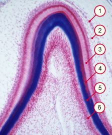
| Root formation takes place only after the formation of the tooth crown is largely completed. In the fold, between the inner and outer enamel epithelium no enamel is produced – the two layers lie closely on one another. This region is also known as Hertwig's epithelial sheath. Via proliferation the enamel organ grows further into the depths in this region and preforms the anlage of the later dental root in that the edges grow conically towards each other and leave one, two or three root canals open. |
|
Fig. 15 - Hertwig's epithelial sheath |
|
Legend |
|
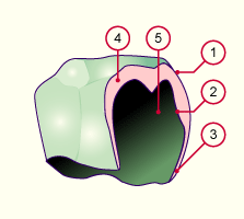
1
2
3
4
5
|
Outer enamel epithelium
Inner enamel epithelium
Hertwig's epithelial sheath
Enamel pulp
Tooth pulp |
|
|
|
Fig. 15
At its edges, the enamel organ grows into the depths and forms the Hertwig's epithelial sheath, which will close up the tooth below except for a root canal (or possibly two or three root canals.
|
|
More info
|
|
Root formation takes place only after the crown formation has been completed. The transition from outer to inner layer of the enamel organ lengthens and forms one, two or three root canals.
|
|
|
| Root formation |
|
Legend |
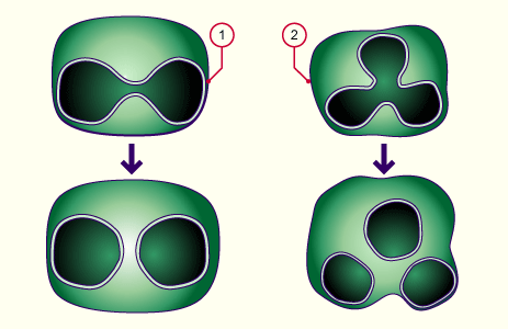
1
2
|
Tooth with two root canals
Tooth with three root canals |
|
|
|
The root canals arise through extension and later fusion of the enveloping layer between the outer and inner enamel epithelium.
|
|
Development of the salivary glands
|
|
|
|
The salivary glands arise only in the later embryonic period (stage 18, ca. 44 days, 18). The first sign is a thickening of the epithelium on the side of the tongue, outside the anlage of the dental arch (lamina dentalis).
In the labiogingival sulcus, on the exterior of the labiodental lamina, arises the anlage of the ectodermal parotid gland (serous glands).
In the linguogingival sulcus that lies towards the tongue, the sublingual (mucous) and submandibular (sero-mucous) glands arise, which have an endodermal origin. They arise in the hyoid arch region.
|
|
|
|
More info
|
|
Since the submandibular gland does not remain on the mouth floor, but in the course of the further development grows below the mandibular arch and the mylohyoid muscle, its outlet crosses over the lingual nerve, which grows into the tongue on both sides.
|
|
|
|
|
|
|