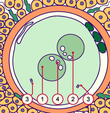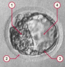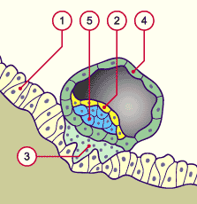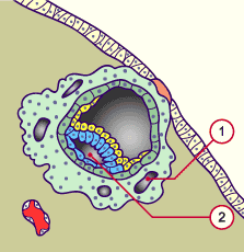
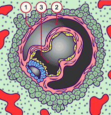
1
2
3 |
Extraembryonic mesoblast
Amniotic cavity
Primary yolk sac
(= primary umbilical vesicle) |
|
|
|
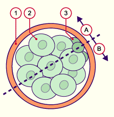
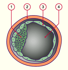
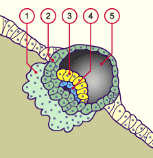
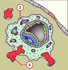
1
2
3 |
Extraembryonic mesoblast
Chorion cavity
Secundary yolk sac
(= secundary umbilical vesicle) |
|
|
|
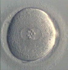
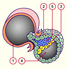
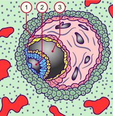
Characteristic signs:
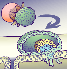
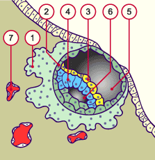
Erosion of the maternal vessels: maternal blood in the lacunae of the throphoblast
The prechordal plate is formed (see stage 6)
Extraembryonic mesoblast
Transformation of the primary into the secundary yolk sac (= secundary umbilical vesicle)
|